
NA • 321974
| Product name | B-Tg(Luc) HCT 116 |
|---|---|
| Catalog number | 321974 |
| Strain name | NA |
| Aliases | NA |
| Tissue | Colon |
| Disease | Colon carcinoma |
| Species | Human |
| Application | B-Tg(Luc) HCT 116 |
This B-Tg(Luc) HCT 116 cell line expresses firefly luciferase as a marker of HCT 116 cells. Fluorescence could be detected in B-Tg(Luc) HCT 116 cells.
Gene targeting strategy for B-Tg(Luc) HCT 116 cells. The exogenous promoter and luciferase coding sequence were inserted into the mouse genome randomly.
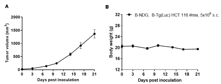
Subcutaneous homograft tumor growth of B-Tg(Luc) HCT 116 cells. B-Tg(Luc) HCT 116 cells (5x10⁶) were subcutaneously implanted into B-NDG mice (female, 8-week-old, n=5). Tumor volume and body weight were measured twice a week. (A) Average tumor volume ± SEM. (B) Body weight (Mean ± SEM). Volume was expressed in mm3 using the formula: V=0.5 X long diameter X short diameter2. As shown in panel A, B-Tg(Luc) HCT 116 cells were able to establish tumors in vivo and can be used for efficacy studies.
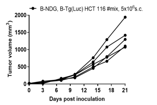
B-Tg(Luc) HCT 116 tumor growth of individual mice. B-Tg(Luc) HCT 116 cells (5x10⁶) were subcutaneously implanted into B-NDG mice (female, 8-week-old, n=5). As shown in panel, B-Tg(Luc) HCT 116 cells were able to establish tumors in vivo and can be used for efficacy studies.
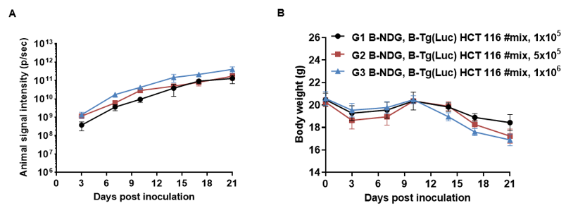
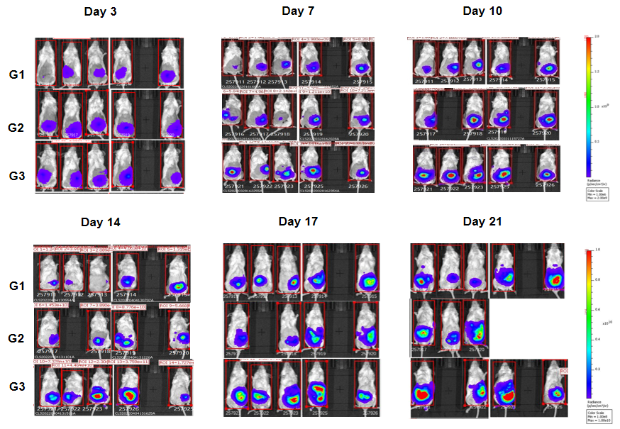
B-Tg(Luc) HCT 116 cells (1x10⁵, 5x10⁵ ,1x106) were injected into the colon of wild-type B-NDG mice. Signal intensity and body weight were measured twice a week. (A) Imaging was performed twice a week. (B) Body weight (Mean ± SEM). B-Tg(Luc) HCT 116 cells can be used for in vivo efficacy evalsuation.
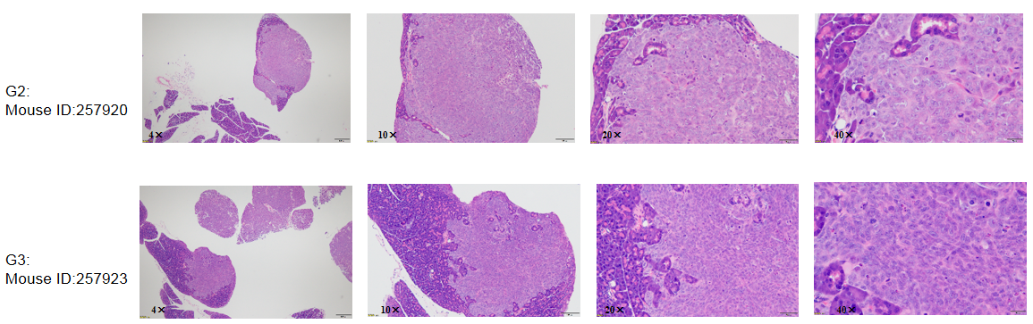
Histopathological HE staining was performed in two suspected tumor metastasis tissue samples.The test results showed that tumor metastasis occurred in the pancreas of mice.