B-hTL1A/hDR3/hFcRn mice

C57BL/6-Tnfsf15tm2(TNFSF15)Bcgen Tnfrsf25tm3(TNFRSF25)Bcgen Fcgrttm1(FCGRT)Bcgen/Bcgen • 114261

B-hTL1A/hDR3/hFcRn mice

Product nameB-hTL1A/hDR3/hFcRn mice
Catalog number114261
Strain nameC57BL/6-Tnfsf15tm2(TNFSF15)Bcgen Tnfrsf25tm3(TNFRSF25)Bcgen Fcgrttm1(FCGRT)Bcgen/Bcgen
Strain backgroundC57BL/6
NCBI gene ID (Human)
AliasesTL1; TL1A; VEGI; TNLG1B; VEGI192A; DR3; TR3; DDR3; LARD; APO-3; TRAMP; WSL-1; WSL-LR; TNFRSF12; FCRN; FcgammaRn; alpha-chain

在此页面上

  • Description
  • Targeting strategy
  • Phenotypic analysis
  • Efficacy

海报

查看全部

    发表文章

      Description

      TL1A: A key inflammation cytokine in chronic intestinal inflammation and fibrosis-related diseases.

      • Gene Information: TNF Superfamily Member 15 (TNFSF15, also known as TL1A) is a protein-coding gene located on chromosome 9q32. This cytokine is a ligand for receptor TNFRSF25 (also known as DR3) and  TNFRSF6B (also known as DcR3).
      • Protein Expression: TL1A is expressed in various immune cells (such as monocytes, macrophages, dendritic cells, and T cells) as well as in non-immune cells (such as synovial fibroblasts and endothelial cells). TL1A is a type II transmembrane protein that exists in either membrane-bound (mTL1A) or soluble (sTL1A) forms.
      • Signaling Pathway: TL1A competes with Death Receptor 3 (DR3) for binding, providing stimulus signals for downstream signaling pathways, thereby regulating the proliferation, activation, apoptosis of effector cells, and the production of cytokines and chemokines.
      • Therapeutic Inhibition: Blocking the interaction between TL1A and DR3 can reduce the severity of autoimmune diseases, such as the IBD model.
      Targeting strategy

      TL1A

      • The exons 1-4 of mouse Tl1a gene that encode extracellular domain were replaced by human counterparts in B-hTL1A/hDR3/hFcRn mice..
      • The genomic region of mouse Tl1a gene that encodes transmembrane domain and cytoplasmic portion was retained. The promoter, 5’UTR and 3’UTR region of the mouse gene were also retained. The TL1A expression was driven by endogenous mouse Tl1a promoter, while mouse Tl1a gene transcription and translation will be disrupted.

      DR3

      • The exons 1-10 of mouse DR3 gene that encode the whole molecule (ATG to STOP codon), including promoter, 5’UTR and 3’UTR were replaced by human counterparts in B-hTL1A/hDR3 mice.
      • The human DR3 expression was driven by human DR3 promoter, while mouse DR3 gene transcription and translation will be disrupted.

      FcRn

      • The human full-length FCGRT cDNA was inserted into the Fcgrt exon 2 of wild-type mice.
      TL1A Protein Expression Analysis
      • Mouse TL1A was detected exclusively in wild-type C57BL/6JNifdc mice
      • Human TL1A was detected in homozygous B-hTL1A/hDR3/hFcRn mice, but not in wild-type mice.

      Strain specific TL1A expression analysis in homozygous B-hTL1A/hDR3/hFcRn mice by ELISA. Bone marrow-derived dendritic cells were isolated from wild-type C57BL/6JNifdc mice (+/+) and homozygous B-hTL1A/hDR3/hFcRn mice (H/H; H/H; H/H) and stimulated with 1 μg/mL LPS in vitro for 24 h, then cell supernatants were collected and analyzed by mouse TL1A ELISA kit and human TL1A ELISA kit (R&D, DY1319-05). Mouse TL1A was exclusively detectable in wild-type C57BL/6JNifdc mice. Human TL1A was exclusively detectable in homozygous B-hTL1A/hDR3/hFcRn mice but not in wild-type mice.

      DR3 Protein Expression Analysis
      • Mouse DR3 was detectable in wild-type C57BL/6JNifdc mice.
      • Human DR3 was exclusively detectable in homozygous B-hTL1A/hDR3/hFcRn mice.

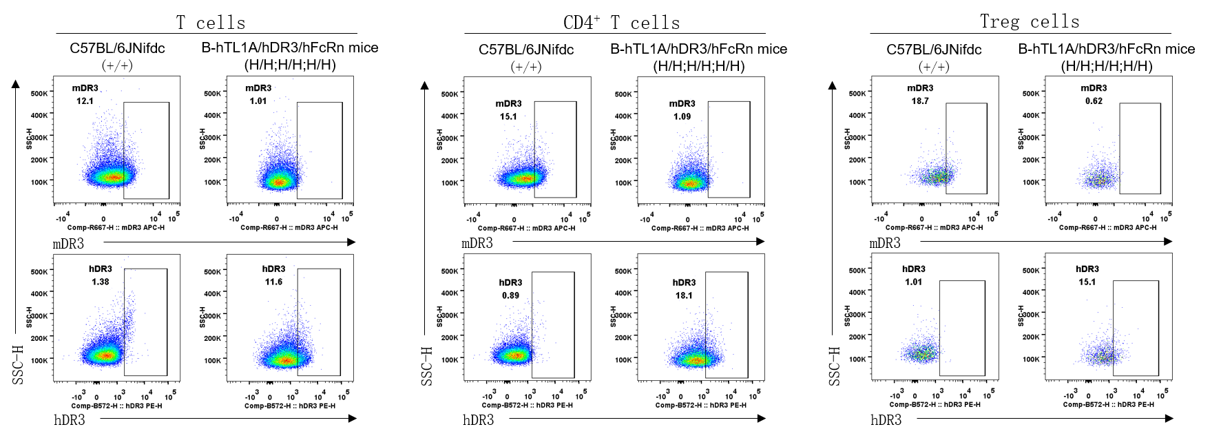
      Strain specific DR3 expression analysis in wild-type C57BL/6JNifdc mice and homozygous B-hTL1A/hDR3/hFcRn mice by flow cytometry. Splenocytes were collected from wild-type C57BL/6JNifdc mice (+/+) and homozygous B-hTL1A/hDR3/hFcRn mice (H/H;H/H;H/H), protein expression was analyzed with anti-mouse DR3 antibody (Biolegend, 144407) and anti-human DR3 antibody (Biolegend, 307105) by flow cytometry. Mouse DR3 was detectable in wild-type C57BL/6JNifdc mice, human DR3 was detectable in homozygous B-hTL1A/hDR3/hFcRn mice.

      FcRn Protein Expression Analysis
      • Mouse FcRn was detectable in wild-type C57BL/6JNifdc mice.
      • Human FcRn was  only detectable in homozygous B-hTL1A/hDR3/hFcRn mice, but not in wild-type mice.

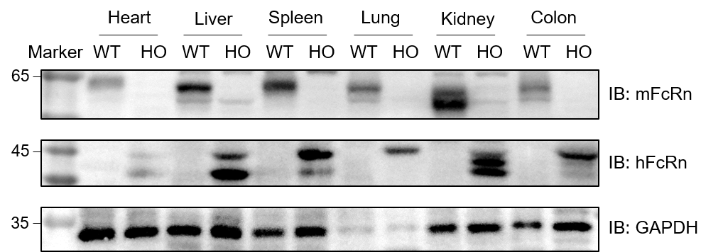
      Western blot analysis of FcRn protein expression in wild-type C57BL/6JNifdc mice and homozygous B-hTL1A/hDR3/hFcRn mice. Various tissue lysates were collected from wild-type C57BL/6JNifdc mice (+/+) and homozygous B-hTL1A/hDR3/hFcRn mice (H/H;H/H;H/H), and then analyzed by western blot with species-specific anti-mouse FcRn antibody (R&D, AF6775) and anti-human FcRn antibody (Novus Biologicals, NBP1-89128). 40 μg total proteins were loaded for western blotting analysis. Mouse FcRn was detectable in wild-type C57BL/6JNifdc mice. Human FcRn was  only detectable in homozygous B-hTL1A/hDR3/hFcRn mice but not in wild-type mice. WT: wild-type C57BL/6JNifdc mice; HO: homozygous B-hTL1A/hDR3/hFcRn mice.

      PK Study of anti-human TL1A antibodies
      • B-hTL1A/hDR3/hFcRn mice could be used as a tool for evalsuating the pharmacokinetics of anti-human TL1A antibodies. 

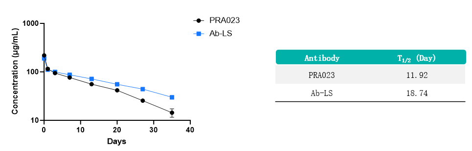
      The pharmacokinetic (PK) study was performed using PRA023 and Ab-LS antibodies in homozygous B-hTL1A/hDR3/hFcRn mice. Mice were intravenously injected with 10 mg/kg PRA023 and Ab-LS antibodies (provided by the client) at Day 0. Blood was collected at designated time points, and serum antibody concentrations were measured. The Ab-LS group exhibited slower clearance compared with PRA023, confirming that the Ab-LS prolonged antibody half-life compared to PRA023 in the B-hTL1A/hDR3/hFcRn mice. Data provided by the client; values expressed as mean ± SEM.

      * When publishing results obtained using this animal model, please acknowledge the source as follows: The animal model [B-hTL1A/hDR3/hFcRn mice] (Cat# 114261) was purchased from Biocytogen.
      站点地图