
C57BL/6-Il11tm1(IL11)Bcgen Il11ratm1(IL11RA)Bcgen/Bcgen • 111865
| Product name | B-hIL11/hIL11RA mice |
|---|---|
| Catalog number | 111865 |
| Strain name | C57BL/6-Il11tm1(IL11)Bcgen Il11ratm1(IL11RA)Bcgen/Bcgen |
| Strain background | C57BL/6 |
| NCBI gene ID | (Human) |
| Aliases | AGIF; IL-11; CRSDA |
Gene targeting strategy for B-hIL11/hIL11RA mice. The exons 1~5 of mouse Il11 gene that encode the full-length protein were replaced by human IL11 exons 1~5 in B-hIL11/hIL11RA mice. The exons 3-11 of mouse Il11ra1 gene that encode the extracellular domain were replaced by human IL11RA exons 3-11 in B-hIL11/hIL11RA mice.
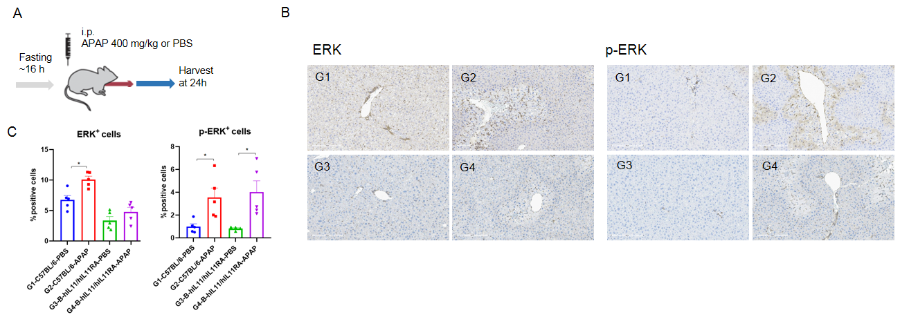
Functional analysis in APAP-induced liver damage in male B-hIL11/hIL11RA mice. (A) Schematic showing the process of APAP-induced liver damage in B-hIL11/hIL11RA mice. B-hIL11/hIL11RA mice (male, 8 weeks, n=5) were fasted overnight and inducted with APAP (400 mg/kg) for 24 h. Liver tissues were collected for functional analysis. (B) Representative IHC images of liver (200 X). (C) Quantification of ERK and p-ERK positive cells from hepatocytes.The IHC results of liver showed that p-ERK increased significantly after APAP induction, and the increase degree was similar to that of wild-type C57BL/6 mice. Values are expressed as mean ± SD. *p<0.05.
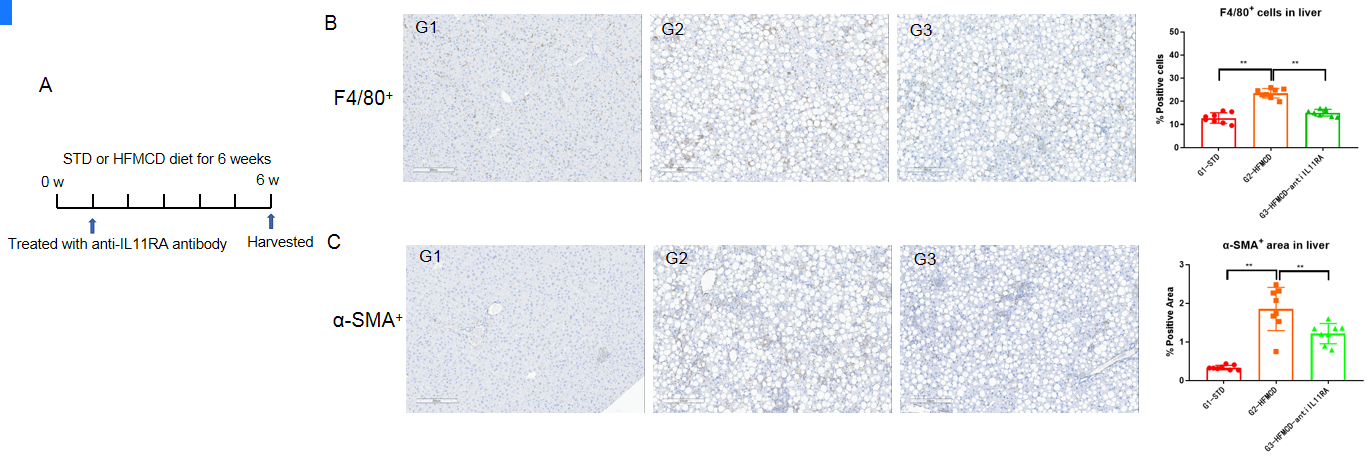
Anti-human IL11RA antibody inhibited the liver damage in male B-hIL11/hIL11RA mice. (A)Schematic showing the process of treatment in B-hIL11/hIL11RA mice. B-hIL11/hIL11RA mice were first randomly separated to receive STD or HFMCD diet for 6 weeks (n=8). The anti-hIL11RA antibody(provided by the client) treatment started when the mice were fed with different diets for 1 week. The liver tissues were collected for analysis. The markers of liver damage, F4/80+ positive cells(B) and α-SMA+ positive areas(C) were increased in the model group(G2) compared to the STD group(G1), indicating that the NASH mouse model was successfully established with B-hIL11/hIL11RA mice. F4/80+ positive cells(B) and α-SMA+ positive areas(C) were reduced in the anti-human IL11RA antibody treated male mice group compared to the model group(G2), indicating that anti-human IL11RA antibody were efficacious in inhibiting liver damage in male B-hIL11/hIL11RA mice. Values are expressed as mean ± SEM. STD, standard diet; HFMCD, high fat methionine and choline deficient diet.